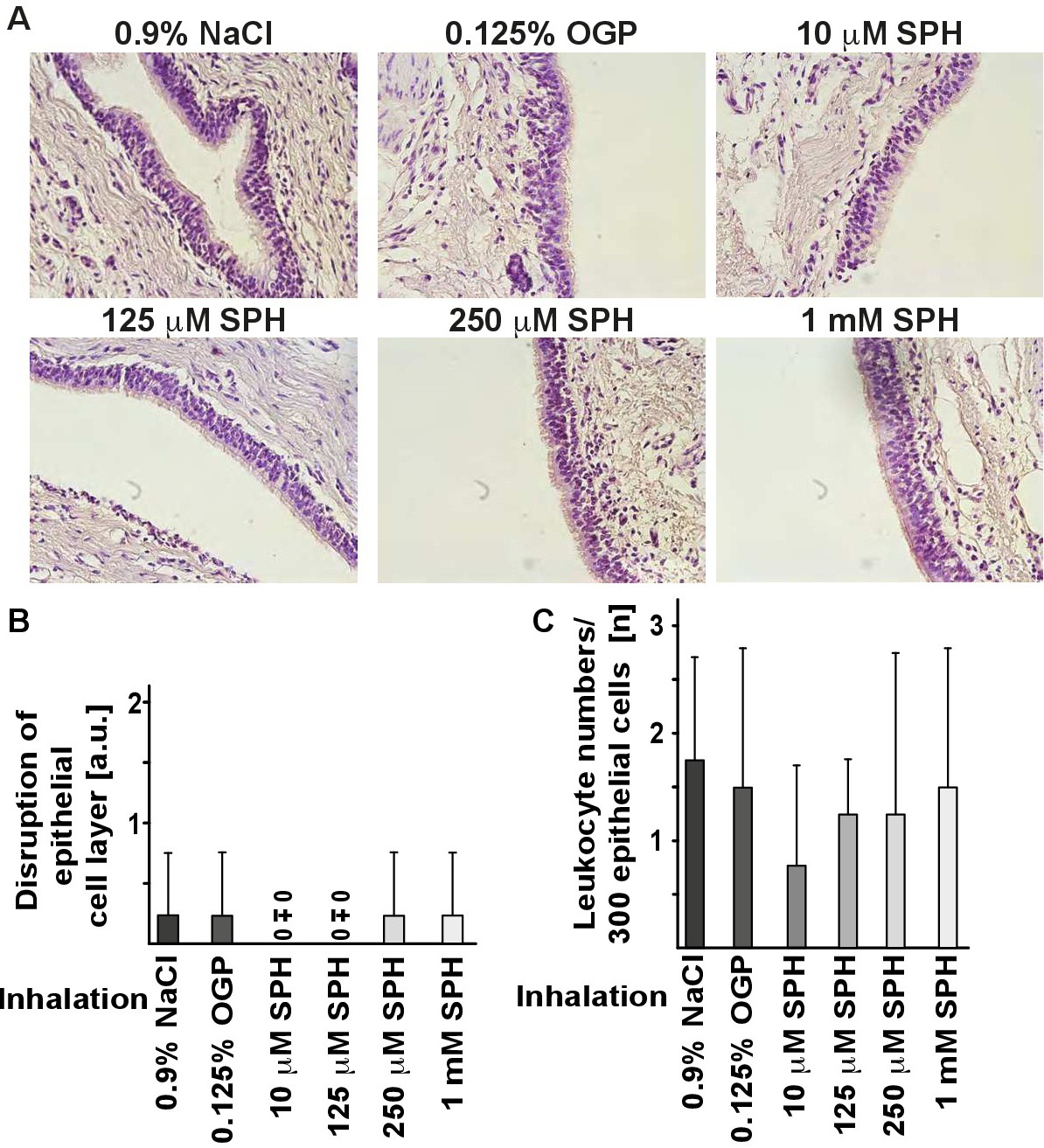
Fig. 4. Inhalation of sphingosine does not affect epithelial cell integrity in bronchi. Paraffin sections from bronchial biopsies from pigs that were inhaled with sphingosine, 0.9% NaCl with 0.125% octylglucopyranoside (OGP) as control or left untreated were stained with hemalaun to analyze the integrity of the bronchial epithelial cell layer and influx of leukocytes into the epithelial cell layer. The studies demonstrated that sphingosine inhalation did not affect epithelial cell integrity (A, B). Sphingosine inhalation also did not induce an influx of leukocytes into the epithelial cell layer (A, C). To determine epithelial cell integrity, we employed the following score: Grade 0: no change of the epithelial cell layer, basal membrane intact, no evidence of leukocyte influx, less than 2% pyknotic, i.e. dead epithelial cells. Grade 1: small disruptions of the epithelial cell layer, basal membrane intact, no evidence of leukocyte influx, less than 5% pyknotic, i.e. dead epithelial cells. Grade 2: Larger disruptions of the epithelial cell layer, basal membrane still intact, no evidence of leukocyte influx, less than 10% pyknotic, i.e. dead epithelial cells. Grade 3: Larger disruptions of the epithelial cell layer, disrupted basal membrane, leukocyte influx, more than 10% pyknotic, i.e. dead epithelial cells. Shown are representative hemalaun stainings from 4 pigs (A) and the quantitative analysis of the epithelial cell integrity (B). In panel C, the number of leukocytes in the epithelial cell layer (total of 300 epithelial cells/pig) was counted. Given are the means ± SD, *p<0.05, **p<0.01, ***p<0.001, ANOVA.